
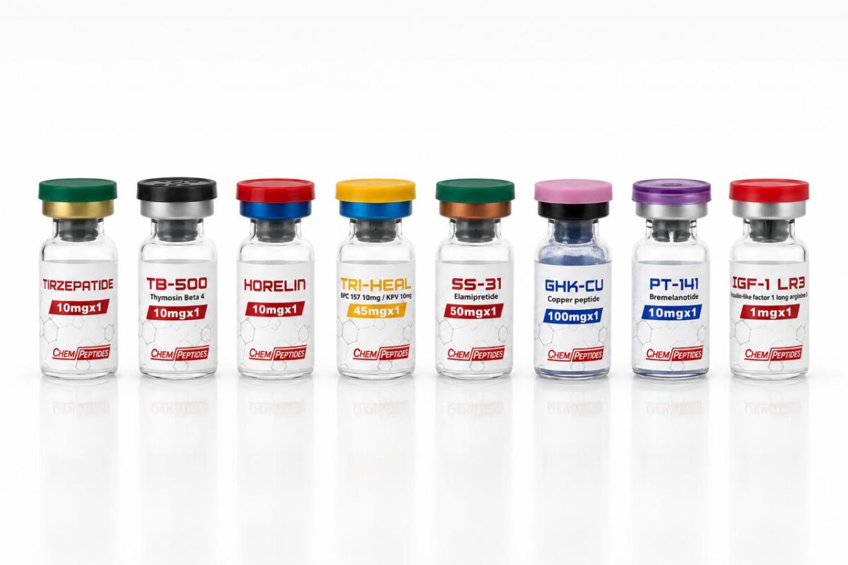
Receptorbindingsassays: Hoe Peptidefarmacologie Wordt Bestudeerd Voordat Er Een Studie Bestaat
Lang voordat een peptide wordt genoemd in een persbericht over een klinische studie, is het gekarakteriseerd in celgebaseerde receptorbindingsexperimenten. Dit zijn de assays die bepalen of een kandidaat-verbinding überhaupt verder komt. Ze worden uitgevoerd in standaard laboratoriumcellijnen, in 96-well- of 384-well-platen, met research-grade peptide als testartikel. Niets eraan vereist een menselijke of dierlijke proefpersoon.
Het Kernprincipe
Een peptidehormoon geeft zijn biologische signaal af door te binden aan een receptor op een doelcel. Meten hoe sterk het bindt, en welk downstream-signaal het activeert, is wat een farmacoloog vertelt of het molecuul de potentie heeft om interessant te zijn. De twee getallen die uit deze experimenten komen zijn EC50 (de concentratie die een halfmaximale respons geeft) en Ki (de inhibitieconstante voor receptorbinding).
Hoe een GLP-1-Receptorassay Daadwerkelijk Wordt Uitgevoerd
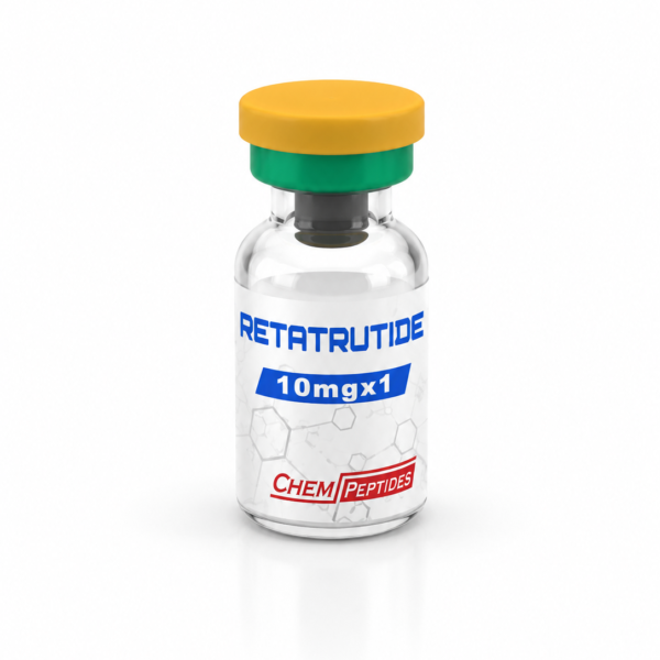
De GLP-1-receptor is een van de meest bestudeerde peptidetargets, omdat hij ten grondslag ligt aan semaglutide, tirzepatide en de nieuwe triple-agonist retatrutide. Een standaard farmacologische workflow:
- Cellijn: HEK293- of CHO-cellen stabiel getransfecteerd met de humane GLP-1-receptor (GLP1R). Deze zijn commercieel verkrijgbaar bij celbanken zoals ATCC.
- Plating: cellen uitgezaaid met 20,000 per well in een 96-well-plaat de dag vóór de assay, in DMEM + 10% FBS.
- Stimulatie: cellen gewassen en vervolgens blootgesteld aan seriële verdunningen van peptide (doorgaans 11 concentraties van 0.01 nM tot 1 µM) gedurende 30 minuten bij 37 °C.
- Uitlezing: cellyse gevolgd door cAMP-kwantificering met een homogene time-resolved fluorescence (HTRF)-kit of AlphaLISA-assay.
- Curve fitting: dosis-responscurves geanalyseerd in GraphPad Prism met vier-parameter logistische regressie. EC50-waarden gerapporteerd met 95% betrouwbaarheidsintervallen.
Coskun en collega’s bij Eli Lilly publiceerden precies dit soort werk voor tirzepatide in Molecular Metabolism (2018, vol. 18, pp. 3-14, “LY3298176, a novel dual GIP and GLP-1 receptor agonist for the treatment of type 2 diabetes mellitus”). Gerapporteerde EC50-waarden: 0.93 nM bij humane GLP-1R en 0.13 nM bij humane GIPR: farmacologie vastgesteld in HEK293-cellen voordat enige klinische studie begon.
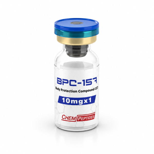
BPC-157 en Celmigratie
Voor een peptide als BPC-157 is de relevante in-vitro assay geen receptorbinding maar celmigratie. De scratch-wound-assay werkt als volgt:
- Fibroblasten (NIH-3T3 of humane dermale fibroblasten) opgekweekt tot confluentie in een 24-well-plaat
- Een pipetpunt door elke well getrokken creëert een uniforme “wond”-opening
- Cellen gewassen en behandeld met BPC-157 bij 1 nM, 10 nM, 100 nM, 1 µM
- Platen in beeld gebracht op 0, 6, 12, 24 uur onder fasecontrastmicroscopie
- Wondsluiting gekwantificeerd in ImageJ als percentage gesloten opening t.o.v. controle
Chang et al. (Journal of Applied Physiology, 2011, 110:774-780) gebruikten deze aanpak om het effect van BPC-157 op migratie en uitgroei van peesafgeleide fibroblasten te karakteriseren. Gepubliceerd, peer-reviewed, in-vitro.
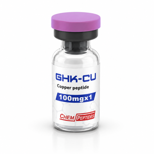
GHK-Cu en Angiogenese
Het tripeptide GHK-Cu (glycyl-L-histidyl-L-lysine gebonden aan koper) wordt bestudeerd op zijn effect op endotheliale buisvorming. De Matrigel-angiogenese-assay:
- HUVEC’s (humane navelstreng-endotheelcellen) uitgezaaid op groeifactor-gereduceerd Matrigel
- Behandeld met GHK-Cu bij 10 nM tot 10 µM, of VEGF als positieve controle
- Buisachtige structuren in beeld gebracht na 8 uur
- Vertakkingspunten en buislengte gekwantificeerd
De reviews van Pickart en Margolina (International Journal of Molecular Sciences, 2018, 19:1987) consolideren decennia in-vitro werk over GHK-Cu en weefselregeneratiepaden op cellulair niveau.
Waarom Dit Werk Echt Wetenschappelijk Gewicht Heeft
Receptorbindings- en celgebaseerde assays zijn de data die peptides in tijdschriften en investeringsdecks krijgen. Een peptide dat een EC50-drempel in HEK293-cellen niet haalt, komt niet verder. Een peptide dat een schone dosis-respons met redelijke potentie laat zien, in twee onafhankelijke assays, is de kandidaat waar een Fase 1-studie omheen wordt ontworpen. Deze filtering vindt volledig plaats in laboratoriumcelkweek: geen mens, geen dier, geen klinische studie.
Wat Onderzoekers Nodig Hebben van een Leverancier
- Geverifieerde identiteit: MS-bevestiging dat het geleverde peptide de geclaimde sequentie is
- Hoge zuiverheid: HPLC ≥ 99% zodat de waargenomen EC50 de geëtiketteerde verbinding weerspiegelt, niet een verontreinigende onzuiverheid
- Batchconsistentie: reproduceerbaarheid van partij tot partij zodat experimentele data doorwerken over replicaatstudies heen
- Documentatie: COA in het dossier voor opname in de methodesectie van elke resulterende publicatie
Dit is wat onderzoekslevering in de praktijk betekent. De flacon die arriveert is gekalibreerd, gekarakteriseerd en klaar voor een 96-well-plaat. Waar het daarna heen gaat, is in een wetenschappelijke dataset: niet in een persoon, niet in een dier.
Uitsluitend voor laboratorium- en analytisch onderzoek. Niet voor menselijke consumptie, dierlijke consumptie of therapeutisch gebruik. Certificates of Analysis per batch beschikbaar op aanvraag.
Gerelateerd onderzoek
- HPLC Zuiverheid van 99 Procent: Waarom Het Verschil Maakt Bij Onderzoek-Grade Peptides
- Wegovy Bijwerkingen: De Complete Gids op Basis van Klinisch Onderzoek 2026
- Analytical Standards in Peptide Research: What Separates a Research Supplier from a Reseller
- HPLC Purity ≥99% in Research Peptides: How High-Performance Liquid Chromatography Verifies Compound Integrity
- Certificate of Analysis (CoA) for Research Peptides: What Every Laboratory Should Verify
Research disclaimer▾
Alle informatie op deze pagina is bedoeld voor laboratoriumonderzoek en in-vitro studies. Geen humane consumptie. Geen medische claims. Producten zijn niet goedgekeurd door EMA of FDA voor therapeutisch gebruik.
Research-grade materiaal bij Chempeptides
De meest gebruikte research peptiden uit onze catalogus:
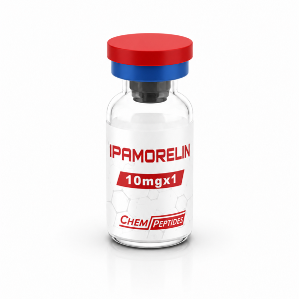
Betalen met iDEAL of Wero. Verzending in discrete verpakking vanuit Nederland. Uitsluitend voor laboratorium- en onderzoeksdoeleinden.